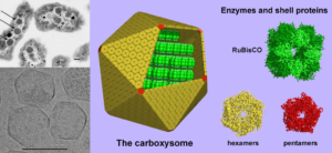
A three-part image of the cyanobacterial structure where carbon dioxide fixation occurs. (Left, above) A thin-section electron micrograph of H. neapolitanus cells with carboxysomes inside. In one of the cells shown, arrows highlight the visible carboxysomes. (Left, below) Purified carboxysomes (material courtesy of S. Heinhorst and G. Cannon) as visualized by cryo-electron microscopy (courtesy of M. Yeager and K. Dryden). (right) Models for the structure of the carboxysome. Current data suggest that the shell is composed of several hundred hexameric protein building blocks and 12 pentameric building blocks. The three-dimensional atomic structures of the shell proteins have been determined by X-ray crystallography. RuBisCO, the main interior enzyme is shown packed inside in a regular arrangement for simplicity, though the actual organization of the enzymes is not understood yet. The other key enzyme, carbonic anhydrase, which is present in lesser amounts, is not illustrated. Scale bars are 100 nm. (image by T. Yeates).
Read the associated commentary:
N. R. Gough, Desert Green Algae and Cyanobacteria for Synthetic Biology. BioSerendipity (7 November 2017).

